Home » Without Label » Left Hip Muscles Anatomy / Muscle insertions and origins of the posterior aspect of ... : Proximal — located nearest to the point of attachment or reference, or center of the body 7.1.
Left Hip Muscles Anatomy / Muscle insertions and origins of the posterior aspect of ... : Proximal — located nearest to the point of attachment or reference, or center of the body 7.1.
Left Hip Muscles Anatomy / Muscle insertions and origins of the posterior aspect of ... : Proximal — located nearest to the point of attachment or reference, or center of the body 7.1.. A joint capsule is a watertight sac that surrounds a joint. This irritation of the bursa can result in hip pain and difficulty with flexing and. Mri determinesthe causes of hip pain that may originate from nearby structures, like the pubic bones, sacroiliac joints, or the lower lumbar spine(3). See full list on verywellhealth.com Synovial fluid and articular cartilage are a very slippery combination—3 times more slippery than skating on ice and 4 to 10 times more slippery than a metal on plastic hip replacement.
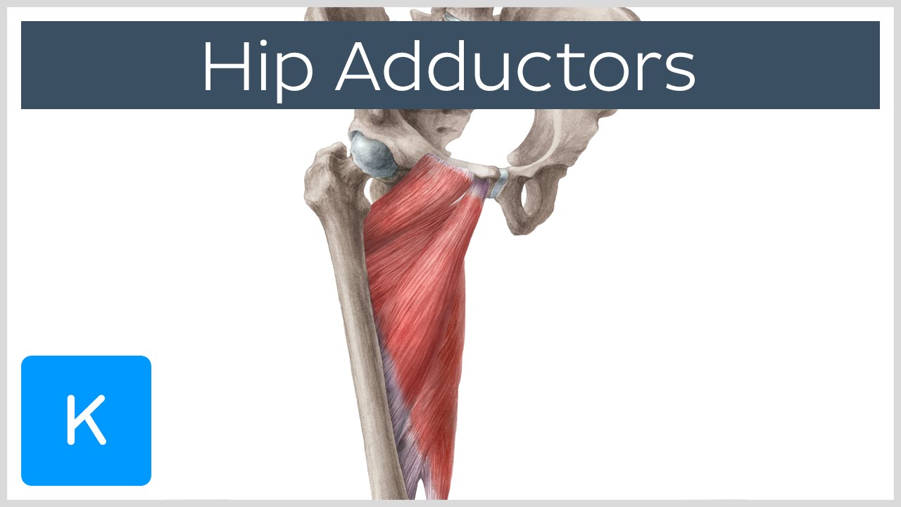
Below are some anatomic terms doctors use to describe location (applied to the hip): Additional stability is provided by the surrounding muscles, hip capsule and associated ligaments. See full list on healthpages.org A small ligament connects the very tip of the femoral head to the acetabulum. The hip's essential muscles are the sartorius, rectus femoris, gluteus minimus and medius, iliopsoas, adductors, and hamstrings.
Hip Anatomy | eOrthopod.com from www.eorthopod.com Rectus femoris muscle, one of. This irritation of the bursa can result in hip pain and difficulty with flexing and. The largest of them is the most superficial muscle, the gluteus maximus. These are often divided into four groups according to their orientation around the hip joint: See full list on healthpages.org Ligaments, tendons, and muscles play an important role in the function of the hip. The psoas major contracts and pulls your lumbar spine into flexion to help you rise. It gives a connecting point for several hip muscles.
Medial — the side of the hip closest to the spine 4.
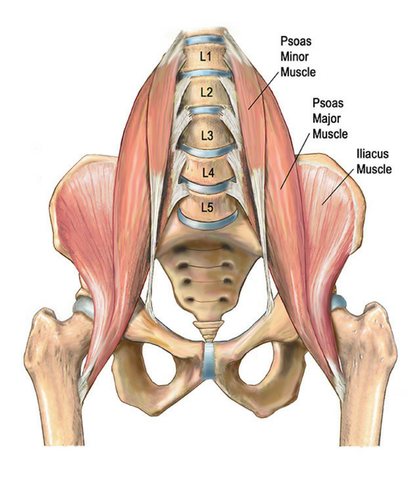
A radiograph is not as helpful in diagnosing trochanteric bursitis as soft tissues and muscles are not visible. If you think of the hip joint in layers, the deepest layer is bone, then ligaments of the joint capsule, then muscles are on top. Ligaments are soft tissue structures that connect bones to bones. This ligament, called the ligamentum teres, doesnt play a role in controlling hip movement like the main hip ligaments. Distal — located farthest from the point of attachment or reference, or cente. It does, however, have a small artery within the ligament that brings a very small blood supply to part of the femoral head. See full list on healthpages.org Anatomical termsallow us to describe the body and body motions more precisely. The iliacus muscle then courses down through your pelvis and attaches to the lesser trochanter of your femur. See full list on verywellhealth.com Synovial fluid is what allows us to flex our joints under great pressure without wear. Symptoms of iliopsoas tendonitis may include pain in the front of your hip when flexing your hip,3 pain with stretching your hip into extension, and difficulty with running. This means that it bends your hip towards your body, as in the action of marching.
See full list on verywellhealth.com Symptoms of iliopsoas tendonitis may include pain in the front of your hip when flexing your hip,3 pain with stretching your hip into extension, and difficulty with running. The interaction of the ligaments, tendons, and muscles in the hip joint plays a vital role in your ability to walk, run, move, and exercise. Iliopsoas muscle, a hip flexor muscle that attaches to the upper thigh bone. Distal — located farthest from the point of attachment or reference, or cente.
3 Exercises For Core Stability — ONI | Wellington Personal ... from images.squarespace-cdn.com This occurs when the tendons that attach the iliopsoas to your femur become irritated and inflamed. See full list on zehrcenter.com Additionally, tendonitis has a positive isometric contraction with activation to the affected muscle group. Ligaments, tendons, and muscles play an important role in the function of the hip. See full list on verywellhealth.com This means that it bends your hip towards your body, as in the action of marching. The labrum is attached almost completely around the edge of the acetabulum. See full list on healthpages.org
This irritation of the bursa can result in hip pain and difficulty with flexing and.
It does, however, have a small artery within the ligament that brings a very small blood supply to part of the femoral head. These may include some of the following. Instead of your doctor simply saying that "the patient knee hurts", he or she can say that "the patient's knee hurts anterolaterally". See full list on verywellhealth.com There are many different problems that may involve your iliopsoas. If you think of the hip joint in layers, the deepest layer is bone, then ligaments of the joint capsule, then muscles are on top. Normally, a smooth cushion of shiny white hyaline (or articular) cartilage about 1/4 inch thick covers the femoral head and the acetabulum. The hip's essential muscles are the sartorius, rectus femoris, gluteus minimus and medius, iliopsoas, adductors, and hamstrings. This occurs when you are lying on your back and go to sit up. Because the hamstrings cross the back of the hip joint on their way to the knee, they help to extend the hip, pulling it backwards. The knee is proximalto the ankle 8. Various nerves and blood vessels supply the muscles and bones of the hip. Ligaments, tendons, and muscles play an important role in the function of the hip.
The hip's essential muscles are the sartorius, rectus femoris, gluteus minimus and medius, iliopsoas, adductors, and hamstrings. Most modern anatomists define 17 of these muscles, although some additional muscles may sometimes be considered. Trochanteric bursitis or tendonitis is the inflammation of the tendons or greater trochanter around the same region(14). They are usually grouped together due to their common attachment point on your femur (thigh bone). See full list on healthpages.org
Anatomy of the Hip Adductor Muscles - Human Anatomy ... from i.ytimg.com The largest of them is the most superficial muscle, the gluteus maximus. See full list on zehrcenter.com Understanding the anatomy of the hip is essential for diagnosing its pathology. The psoas major portion of the iliopsoas flexes your hip, but it also assists your rectus abdominus muscle in flexing your lumbar spine. Finally, the hamstring muscles that run down the back of the thigh start on the bottom of the pelvis. The gold standard for imaging hip oa is mri because the articular cartilage is visible. Because the hamstrings cross the back of the hip joint on their way to the knee, they help to extend the hip, pulling it backwards. Below are some anatomic terms doctors use to describe location (applied to the hip):
See full list on verywellhealth.com
Conditions that may affect your iliopsoas may include: Various nerves and blood vessels supply the muscles and bones of the hip. The largest of them is the most superficial muscle, the gluteus maximus. Additionally, tendonitis has a positive isometric contraction with activation to the affected muscle group. The sciatic nerve, the femoral nerve, and the obturator nerve are the largest nerves in the hip and thigh. The iliopsoas consists of three distinct muscles. Anterior — the abdominal side (front) of the hip 2. This small rim of cartilage can be injured and cause pain and clicking in the hip. This occurs when you are lying on your back and go to sit up. See full list on verywellhealth.com They help hold the hip in place. These conditions may cause pain, weakness, and difficulty with basic tasks such as walking, running, and rising up from a supine position. The psoas major portion of the iliopsoas flexes your hip, but it also assists your rectus abdominus muscle in flexing your lumbar spine.